This Egyptian mummy is called the Gilded Lady for her gold face mask.

© THE FIELD MUSEUM, IMAGE NO. A115214D_030B, CAT. NO. 30007, PHOTOGRAPHER JOHN WEINSTEIN

STANDARDS

NGSS: Core Idea: PS4.C

CCSS: Reading Informational Text: 5

TEKS: Science: 3.6A, 4.6A, 5.6A, 3.2A, 4.2A, 5.2A, 6.2A; ELA: 3.3, 4.1, 5.1, 6.1

Mummy Vision!

X-rays show ancient mummies in a new light

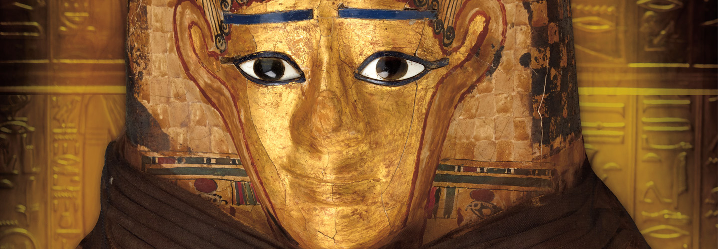
Last year, a 14-year-old boy named Minirdis (mihn-ER-diss) took a road trip across the U.S. But instead of admiring the view, he rode in a box in the back of a truck. That’s because Minirdis is no ordinary boy. He’s a mummy! 

Minirdis was the son of a priest who lived 2,500 years ago in Egypt. When the boy died, his body was mummified, or preserved and wrapped in cloth (see How to Make a Mummy). Then it was placed in a decorated coffin and buried along the Nile River, where it was discovered in the late 1800s.  

The mummy of a 14-year-old boy took a road trip across the U.S. last year. His name was Minirdis (mihn-ER-diss). He lived 2,500 years ago in Egypt.

Minirdis was the son of a priest. When the boy died, his body was mummified. It was preserved and wrapped in cloth (see How to Make a Mummy). Then his mummy was placed in a decorated coffin and buried along the Nile River. It was discovered in the late 1800s.

In 1925, Minirdis’s mummy was donated to the Field Museum in Chicago. For 90 years, it sat in storage with other artifacts the museum didn’t have room to display. 

Now Minirdis is finally on view. He’s one of 20 mummies in a traveling exhibit called “Mummies: Images of the Afterlife.” After stays in two other cities, the exhibit is opening in Denver this month.

Mummies are delicate, and moving them is risky. Harsh temperatures or a bumpy ride could easily damage the ancient remains. Before the trip, conservators at the Field Museum had to examine Minirdis and the other mummies to make sure they were fit for the journey. Plus, they were curious: What was inside the coffins that had been sealed for thousands of years?

Minirdis’s mummy was donated in 1925 to the Field Museum in Chicago, Illinois. The museum houses historic artifacts and items from the natural world. The mummy sat in storage for 90 years with other ancient objects there wasn’t room to display. 

Now Minirdis is finally on view. He’s one of 20 mummies in a traveling exhibit. It’s called “Mummies: Images of the Afterlife.” The exhibit has visited two cities so far. It opens in Denver, Colorado, this month. 

Mummies are delicate. Moving them is risky. Harsh temperatures or a bumpy ride could easily damage the ancient remains. Conservators care for objects in museums. They examined Minirdis and the other mummies before the trip. They needed to make sure the mummies were fit for the journey. Plus, they were curious what was inside the coffins. They’d been sealed for thousands of years!

Seeing Inside

The conservators didn’t want to open the coffins and risk harming the mummies inside. So they turned to a machine called a CT scanner. This device creates 3-D images using X-rays—the same kind that doctors use to look at broken bones. 

X-rays are a form of radiation that can pass through coffins and soft tissue such as skin and muscle. The rays bounce off denser material like bone. This creates an image of the body’s inner layers. A CT scanner shoots X-rays from many angles to create a three-dimensional picture of an object’s insides. 

People have to stay still for CT scans or the images come out blurry. But mummies make perfect patients. They don’t move a muscle! That means scientists can get very detailed images, says J.P. Brown. He’s a conservator at the Field Museum who prepared the mummies for their trip.

The conservators didn’t want to open the coffins. That would risk harming the mummies inside. So they turned to a machine called a CT scanner. This device creates 3-D images using X-rays. X-rays are a form of radiation, or invisible energy. Doctors also use them to look at broken bones. 

X-rays can pass through coffins. The rays also pass through soft tissue such as skin and muscle. But X-rays bounce off thicker material like bone. This reveals an image of the body’s inner layers. A CT scanner shoots X-rays from many angles. This creates a 3-D picture of an object’s insides. 

People have to stay still for CT scans or the images come out blurry. But mummies make perfect patients. They don’t move a muscle! That means scientists can get detailed images, says J.P. Brown. He’s a conservator at the Field Museum. He prepared the mummies for their trip.

Mummies Unwrapped

To scan a mummy, Brown and his team gently place the coffin on a platform that slides into the CT scanner. As the mummy moves through the cylindrical machine, X-rays hit it from all sides (see Inside a Scanner). Over five days, the scientists scanned seven mummies—almost. The bottom section of one coffin was too tall to fit into the machine. The mummy’s feet couldn’t be scanned! 

The scans revealed details about the people in the coffins. By studying a mummy’s bone structure, scientists can tell the age and gender of the person inside. The images also showed clothes and jewelry the mummies were buried with. 

One mummy, a 40-year-old woman called the Gilded Lady, had a decorated face mask. A scan showed remnants of curly hair beneath her wrappings. This helped museum staff build a realistic model of how she looked when she was alive.

Brown and his team gently place a coffin on a platform to scan a mummy. It slides into the CT scanner. The mummy moves through the tube-shaped machine. X-rays hit it from all sides (see Inside a Scanner). The scientists scanned seven mummies over five days. But one coffin was too tall for the machine. The mummy’s feet couldn’t be scanned! 

The scans revealed details about the people in the coffins. Scientists could study each mummy’s bone structure. That told them the age and gender of the person inside. Scans also showed clothes and jewelry the mummies were buried with. 

One mummy belonged to a 40-year-old woman with a decorated face mask. She’s nicknamed the Gilded Lady. A scan showed curly hair inside her wrappings. That helped museum staff build a model of the woman. It showed how she looked when she was alive.

©S.ENTRESSANGLE, E.DAYNES/RECONSTRUCTION ELISABETH DAYNES, PARIS/LOOKATSCIENCES

This artist used scans of Minirdis’s mummy to reconstruct his appearance in life.

Emergency Operation

When conservators scanned Minirdis, they saw a lot of damage inside his coffin. “His legs had separated at the knees, and the feet had fallen off,” says Brown. “It was a mess.”

It was clear that they needed to open Minirdis’s coffin to repair him. With the scans as a guide, they carefully lifted the lid. They used a fine vacuum cleaner to remove loose dirt from the mummy. They repositioned his broken parts, then wrapped fresh linen over the old cloth. Now Minirdis was ready to join his fellow mummies on tour.

The mummies are exhibited alongside images from their CT scans. Brown hopes the scans help visitors understand the ancient people’s lives. “Almost everything we’ve learned would have been impossible without this technology,” he says.

Minirdis’s scan showed a lot of damage. “His legs had separated at the knees,” says Brown. “And the feet had fallen off. It was a mess.”

Conservators needed to open Minirdis’s coffin to repair him. They carefully lifted the lid. Then they used a fine vacuum cleaner to remove loose dirt from the mummy. They moved his broken parts back into place. They wrapped fresh linen over the old cloth. Now Minirdis was ready to join his fellow mummies on tour.

The mummies are displayed with images from their scans. Brown hopes the images help visitors learn about the ancient people’s lives. “Almost everything we’ve learned would have been impossible without this technology,” he says.

videos (1)
Skills Sheets (3)
Skills Sheets (3)
Skills Sheets (3)
Games (1)
Lesson Plan (2)
Lesson Plan (2)
Text-to-Speech